


Patient examination
Examination of a patient with a traumatic dental injury
A rational examination procedure is essential in order to establish a complete and correct diagnosis of all soft and hard tissue injuries.
Take a short medical and dental history
The medical history should include the patient’s general health, allergies and other information that may influence treatment. Be alert to other injuries and symptoms which can indicate more serious injury. It is also of value to ask what has been done before the patient reached the clinic, such as any treatment carried out elsewhere and how avulsed teeth have been stored. The dental history should indicate previous dental traumas, information which may explain radiographic findings such as pulp canal obliteration or apical pathology.
Questions relating to the injury
- Where did the injury occur? This information may have legal implication for the patient and may on occasion indicate the possibility of contamination.
- How did the injury occur? This may lead to identification of the impact zones, i.e. a chin injury is often combined with crown or crown-root fractures in premolar and molar regions.
- When did the injury occur? This information may be essential in relation to many injury types. In relation to a tooth avulsion the extent of time and the extraoral storage condition becomes very decisive for later treatment.
- Was there a period of unconsciousness? If so, for how long? Amnesia, nausea and vomiting are all signs of brain damage and require medical attention.
- Is there any disturbance in the bite? An affirmative answer may indicate a luxation injury or root fracture with displacement, an alveolar or jaw fracture or a fracture of the condylar region.
- Is there any reaction in the teeth to cold and/or heat exposure? A positive finding indicates exposed dentin and/or pulp.
Clinical examination
A systematic approach is recommended. The general principle with oral injuries to “examine from outside to inside and treat from inside to outside” is helpful for a systematic approach. Hence suturing of lip lacerations should be carried out after intraoral injuries of urgent character have been treated. Otherwise it may be difficult to enter the oral cavity once edema has started to develop.
- Clean the face and the oral cavity with water or saline
If there are soft tissue wounds, a mild detergent should be used. This cleaning will make the patient feel more comfortable and facilitate extraoral and oral examination. - Examine the face, lips and oral muscles for soft tissue lesions.
- Palpate the facial skeleton for signs of fractures.
- Inspect the dental trauma region for fractures, abnormal tooth position, tooth mobility and abnormal response to percussion. Furthermore, registration of direction of displacement in case of luxation injuries. In case of fractures their extent and involvement of dentin, pulp and the relation to the gingival sulcus area is noted.
- Pulp testing (usually electrometric) completes the clinical examination.
- Percussion testing of teeth may reveal a high pitch bony sound indicating intrusion of the tooth.
- The possibility of inhaled or swallowed teeth, parts of teeth or prosthetic appliances at the time of injury should always be considered. Foreign body in the airways usually give symptoms such as coughing but may go undetected, which is especially important when treating unconscious patients.
Radiographic examination
The completed clinical examination has now identified the trauma region and this site should now be examined with relevant radiographic techniques. Several clinical studies have shown that multiple radiographic procedures are needed to detect displacement of the tooth in its socket as well as presence of root fractures It is essential to consider the radiographic film format used in order to achieve a high quality image of the traumatized tooth. A steep occlusal exposure (using a size 2 film (DF 58, EP 21)) of the traumatized anterior region gives an excellent view of most lateral luxations, apical and mid-root fractures and alveolar fractures.
A minimum series of radiographs for a typical dental trauma to the anterior region of the maxilla is: 3 films with parallel technique supplemented with one film with steep occlusal exposure.
In case of lip laceration a radiograph with the film placed inside the lip and reduced exposure time should be added.
Cone beam CT may be a valuable tool and can be considered if the obtained information will influence the choice of treatment. An example could be a tooth with a crown-root fracture where the extend of the fracture will determine whether it will be possible to preserve the tooth or not.
Radiographic examination of soft tissue lesions
In the presence of a penetrating lip lesion, a soft tissue radiograph is indicated in order to locate any foreign bodies. It should be noted that the oral orbicular muscles close tightly around foreign bodies in the lip, making them impossible to palpate; they can only be identified radiographically. This is accomplished by placing a dental film between the lips and the dental arch and using the lowest exposure time. If this exposure reveals foreign bodies (a radiographic examination will normally demonstrate foreign bodies such as tooth fragments, composite filling material, metal, gravel, whereas organic materials such as cloth and wood cannot be seen), a lateral radiograph can be added (at 50% normal exposure time) to visualize the foreign bodies in relation to the cutaneous and mucosal surfaces of the lips. With the combined information from the clinical and radiographic examinations, diagnosis, prognosis and treatment planning can be accomplished.
Supplemental radiographs such as panoramic (OPG) views and Cone beam CT can be of value and considered when indicated and available.
Photographic registration
Finally, photographic registration of the trauma is recommended as it offers an exact documentation of the extent of injury and can be used later in treatment planning, legal claims or clinical research. Note that a patient consent is required before taking such photographs and when considering using them for other purposes such as research and publication.
Diagnosis
The information obtained from clinical and radiographic examination will lead to the correct diagnosis including both fractures and luxation injuries (combination injuries). To help the newcomers in dental traumatology the Trauma Pathfinder can guide the practitioner via a series of ‘yes’ and ‘no’ questions to a correct diagnosis.
Recently, a new index based on 5 digits has been developed which comprise all important information which can be easily computerized. This index enables combination injuries to be registered at the same time (Eden Baysal index). Standardized registration will facilitate comparison of data and outcome between different centers worldwide enabling larger materials for research.
Modified Eden Baysal Dental Trauma Index
| Number of tooth (FDI) | In parenthesis | ||
| 1st digit Crown fracture |
0 to 5 | 0 = none 1 = enamel fracture 2 = enamel and dentin fracture = uncomplicated crown fracture 3 = enamel and dentin fracture including the pulp = complicated crown fracture 4 = enamel-dentin-cementum fracture = uncomplicated crown-froot fracture 5 = Enamel-dentin-cementum fracture including the pulp = complicated crown-root fracture |
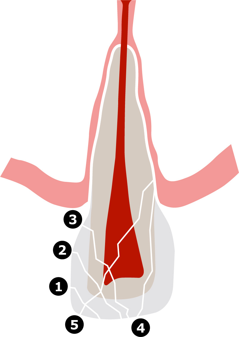
(Click image for detailed figure) |
| 2nd digit Root fracture |
0 to 3 | 0 = none 1 = apical 1/3 root fracture 2 = middle 1/3 root fracture 3 = cervical 1/3 root fracture |
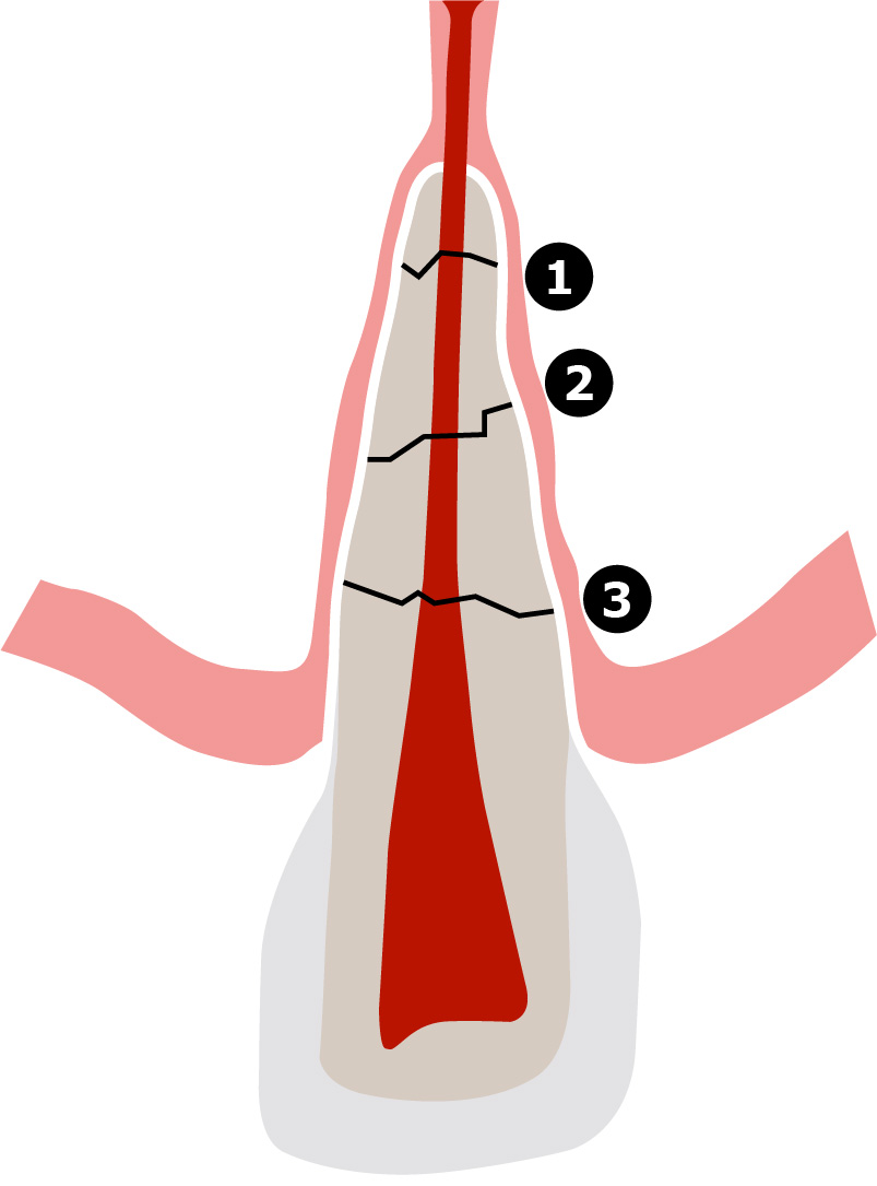
(Click image for detailed figure) |
| 3rd digit Luxation injury |
Capital first letters |
N = None C = Concussion S = Subluxation L = Lateral luxation E = Extrusive luxation I = Intrusive luxation A = Avulsion |
|
| 4th digit Maturity and shape of apex |
Small first letters |
i = immature m = mature r = resorbed |
|
| 5th digit Alveolar process fracture |
+ or – | + sign = Alveolar process fracture – sign = No alveolar fracture |
|
| Soft tissue injury | Superscript 0 to 8 |
0 = none 1 = skin/lip abrasion 2 = skin/lip laceration 3 = skin/lip contusion 4 = skin/lip avulsion 5 = intra-oral abrasion 6 = intra-oral laceration 7 = intra-oral contusion 8 = intra-oral avulsion |
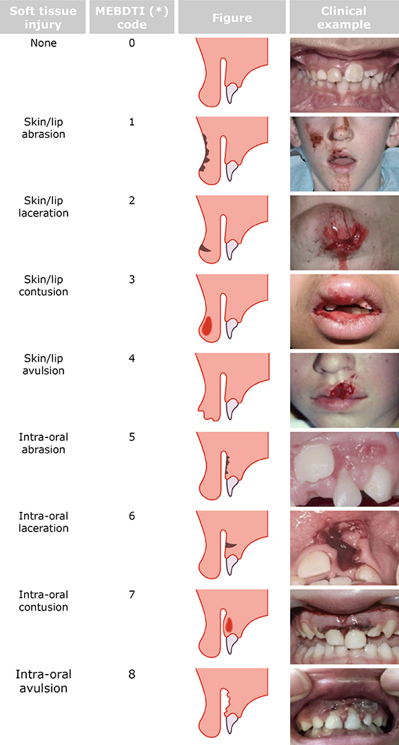 (Click image for detailed figure) |
Recording Baseline Diagnostic Findings
To generate codes with ‘Modified Eden Baysal Dental Trauma Index’ please click the link below:
Generator
For more information see manual
Eden Baysal Dental Trauma Outcome Index
| Number of tooth (FDI) | In parenthesis | ||
| 1st digit Crown restoration |
0 to 9 | 0 = no crown fracture = need for restoration 1 = intact fragment reattachment 2 = acceptable restoration 3 = discoloration of the restoration – no renewal necessary 4 = renewal necessary 5 = restoration missing 8 = not clear 9 = tooth missing |
|
| 2nd digit Root fracture healing |
0 to 9 | 0 = no root fracture 1 = hard tissue healing 2 = connective tissue healing with bone 3 = no healing and infection present 8 = not clear 9 = tooth missing |
|
| 3rd digit External root resortion type |
Capital letters |
NI = No injury of PDL PH = Proper healing of PDL IR = Inflammatory resorption (infection-related resorption) RR = Replacement resorption (ankylosis-related resorption) 8 = not clear 9 = tooth missing |
|
| 4th digit Root development |
Small letters |
m = mature root i = immature growing root a = arrested root development r = resorbed root (physiological) 8 = not clear 9 = tooth missing |
|
| 5th digit Pulp status |
0 to 9 | 0 = clinically normal pulp 1 = partial / total pulp canal calcification 2 = vital endodontic / regenerative treatment 3 = necrotic / infected pulp 4 = root canal treatment performed 8 = not clear 9 = tooth missing |
To generate codes with ‘Eden Dental Trauma Outcome Index’ please click the link below:
Generator
For more information see manual
Recommended reading
1. ANDREASEN FM, ANDREASEN JO. Diagnosis of luxation injuries: the importance of standardized clinical, radiographic and photographic techniques in clinical investigations. Endod Dent Traumatol 1985;5:160-169.
2. BAKLAND LK, ANDREASEN JO. Examination of the dentally traumatized patient. Calif Dent Ass J 1996;24:35-44.
3. ANDREASEN FM, ANDREASEN JO, TSUKIBOSHI M, COHENCA N. Examination and Diagnosis of Dental Injuries. In: Andreasen JO, Andreasen FM, Andersson L, (eds.). Textbook and Color Atlas of Traumatic Injuries to the Teeth (5th ed.). Oxford, Blackwell 2019, pp. 295-326
4. EDEN E, BAYSAL M, ANDERSSON L. Eden Baysal Dental Trauma Index: Face and content validation. Dent Traumatol. 2020;36:117–123.
5. EDEN E, ONETTO JE, O’CONNELL AC. Extension of a novel diagnostic index to include soft tissue injuries: Modified Eden Baysal Dental Trauma Index. Dent Traumatol. 2021;37:749-757.
6. KAVAL ME, ATEŞÇI AA, KURT SSM, BOYACIOĞLU H, EDEN E. Assessment of the validity and reliability of a novel dental trauma index. Dent Traumatol. 2023;39:381-385.
7. EDEN E, ONETTO JE, LAURIDSEN E, ANDERSSON L. Simplified Registration of Clinician-Important Traumatic Dental Injury Outcomes Using a New Index. Dent Traumatol. 2025 Jul 16. doi: 10.1111/edt.13086. Epub ahead of print. PMID: 40665855.